The DNA backbone
Nucleotides within a DNA strand are joined together by strong covalent bonds located in the DNA backbone. The chemical elements in the backbone are responsible for many of the physical properties of DNA, such as charge and strength. In this activity, you will explore features of the backbone and learn about the bonds that hold nucleotides together.
Worksheets
The DNA backbone and the double-strand DNA drawing
Instructions
1. Open the DNA structure file, 1NAJ.cn3. The backbone of each DNA strand is made of sugar residues that are held together with phosphodiester bonds. To see the backbones more clearly, open the Style menu. Select Edit Global Style.
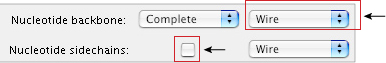
2. Change the settings for the Nucleotide backbone to show: Complete, Ball and Stick, and Element, as shown in the Style Options window above.
3. Change the settings for the Nucleotide sidechains to show Space Fill and Element (The pull-down menu in the image on the right shows "Molecule." this should be changed to element). Make sure that the box in the Show column is checked.
4. Click Apply > Done.
5. The backbone for each DNA strand now appears on the outside of the DNA, with the bases in the middle. Turn the DNA so that you're viewing it from one end with the backbone along the outside and the bases in the center. Look at the colors, and use your color key, from the DNA building block activity, to locate the different elements.
a. Which element appears in the backbone, but not in the bases?
b. Which element appears in the center, but not in the backbone?
6. To see the backbone structures more clearly, remove the bases. Open the Style menu. Select Rendering Shortcuts > Toggle Sidechains.
Only the backbones of the two DNA strands are visible now. The DNA backbone is referred to as the "sugar-phosphate" backbone because it contains deoxyribose groups (the sugars), held together with phosphodiester bonds (each phosphodiester bond contains one phosphate group).
7. If you've moved the DNA structure, turn it back again so that you’re looking through the center. Notice where the phosphate groups are located. What charge do the phosphate groups give to the outside of the DNA molecule?
8. Change the view to show only one strand. To do this, use the pointer to select all of the bases in either strand of DNA in the Sequence/Alignment Viewer window.
9. Open the Select menu and choose Show Selected Residues.
10. Click the pointer in the Sequence/Alignment Viewer window to deselect the sequence and see it colored by element.
11. Compare the sketch of double-stranded DNA (at the end of your worksheet handout), in the DNA worksheet, to the structure shown in Cn3D. The sugar phosphate backbone has sugar rings, each with 5 carbons and an oxygen, that are held together by phosphodiester bonds.
Find, circle, and label examples of each of the chemical groups or atoms, listed below.
a. a deoxyribose
b. a phosphate
c. a phosphorus
d. a 5' carbon at the end of a DNA strand
e. a 3' hydroxyl (OH) group at the end of a DNA strand
f. a 3' carbon
12. Which of the groups listed above gives DNA its negative charge?
14. Open the Style menu and select Edit Global Style. Now, we are going to remove the hydrogens to get a better view of the sugar phosphate backbone.
a. Change the rendering style for the Nucleotide backbone to Wire.
b. Deselect the check box next to Nucleotide sidechains to make them invisible.
c. Deselect the checkbox next to Hydrogens to quit showing the hydrogens.
15. Click Apply > Done.
Look at the bonds holding the nucleotides together. There will be a green colored atom, attached to the sugars by red colored atoms on each side. These types of bonds are called "phosphodiester bonds." An ester bond is a bond where oxygen is bound to two different elements, other than hydrogens, in this form element-O-element.
The prefix "di" means "two." A diester bond has two ester bonds. In this case the bonds are arranged like this:
C-O-P-O-C. Phosphodiester bonds are the covalent bonds that hold nucleotides together in a single strand of DNA.
a. Find a phosphodiester bond in the three-dimensional structure. Then, find, circle, and label a phosphodiester bond in the drawing of double-stranded DNA.
b. Find, circle, and label a single ester bond on the worksheet.
16. Select Style > Edit Global Style. Click the check box next to Nucleotide sidechain to see the bases reappear. Change the rendering of the these side chains to Wire. Click Done.
17. Turn the structure so that you’re viewing the bases sideways as flat planes. How are the sugars oriented relative to the bases? Describe how this looks.
18. Open the Select menu and choose Show Everything to show both strands of DNA.
19. Look at two sugars on opposite strands. Are the sugars from the two strands pointed in the same direction?
If you're having a hard time determining the orientation of the sugars, look for the oxygen atoms within the sugar rings.